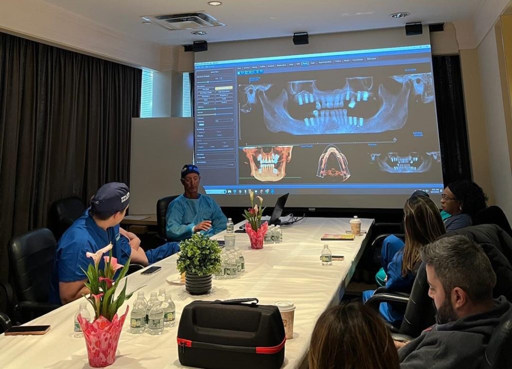
Cone beam computed tomography (CBCT) gives general dentists a precise, three-dimensional view of bone volume, density, and critical anatomical structures before placing a dental implant. Unlike conventional two-dimensional periapical or panoramic radiographs, CBCT captures the full spatial relationship between the proposed implant site and structures such as the inferior alveolar nerve, maxillary sinus, and adjacent tooth roots. When used as part of a thorough treatment-planning process, CBCT imaging reduces the risk of surgical complications and supports more predictable implant outcomes.
If you are exploring how to choose the right implant training program that incorporates modern imaging technology, understanding CBCT is an essential first step.
What Is CBCT Imaging and How Does It Differ from Conventional Dental X-Rays?
Cone beam computed tomography is a radiographic technique that rotates an X-ray source and a flat-panel detector around the patient’s head, capturing hundreds of individual projection images. Software then reconstructs those projections into a three-dimensional volumetric dataset that the clinician can view in axial, coronal, sagittal, and cross-sectional planes. The entire acquisition typically takes 10 to 30 seconds of beam-on time at the chairside unit.
Conventional periapical and panoramic radiographs display anatomy in only two dimensions, which inevitably produces geometric distortion and overlapping structures. CBCT significantly reduces that overlap by rendering each plane independently. The American Academy of Oral and Maxillofacial Radiology (AAOMR) published a position statement in 2012 identifying CBCT as the imaging method of choice for cross-sectional assessment of dental implant sites. The AAOMR recommends that initial radiographic evaluation begin with a panoramic radiograph, with CBCT reserved for cases where additional cross-sectional information is needed to evaluate bone dimensions or proximity to vital structures. [1]
For the general dentist, the practical implication is significant. A panoramic radiograph may suggest adequate bone height above the inferior alveolar canal, but it cannot accurately convey buccolingual width or cortical thickness. CBCT provides both, allowing the clinician to determine whether a site can accommodate a standard-diameter implant without augmentation or whether a bone grafting procedure is required before surgical placement.
Why CBCT Is Considered the Gold Standard for Pre-Implant Assessment
The clinical case for CBCT in implant dentistry rests on three pillars: anatomical accuracy, surgical safety, and prosthetically driven planning.
From an anatomical accuracy standpoint, CBCT allows the clinician to measure bone height and width with sub-millimeter precision. A systematic review by Fokas et al. (2018) published in Clinical Oral Implants Research analyzed 22 studies on CBCT linear measurement accuracy for implant planning. CBCT has been validated as a reliable tool for three-dimensional preoperative implant planning, though clinicians should apply a minimum 2 mm safety margin from adjacent anatomic structures to account for potential measurement variability. Real-world accuracy may be reduced by factors including patient movement, metallic artifacts, differences in scanner exposure settings, the planning software used, and whether measurements are taken manually or through automated methods. [2]
Surgical safety is equally well supported in the literature. A 2018 systematic review and meta-analysis by Tahmaseb et al. published in Clinical Oral Implants Research evaluated the accuracy of static computer-aided implant surgery across 2,238 implants in 471 patients. The study found a mean error of 1.2 mm at the entry point, 1.4 mm at the implant apex, and an angular deviation of 3.5 degrees. These findings demonstrate that CBCT-guided placement achieves clinically meaningful precision, and the authors recommended maintaining a minimum 2 mm safety margin from adjacent anatomical structures. [3]
Prosthetically driven planning is the third pillar and arguably the most transformative. When CBCT data is imported into implant planning software, the restorative team can virtually position the implant to optimize emergence profile, interocclusal space, and axial inclination before the patient enters the operatory. The resulting surgical guide translates the virtual plan into an accurate physical template, dramatically reducing intraoperative decision-making. The International Team for Implantology (ITI) has endorsed this approach, noting that CBCT is preferable over conventional CT when cross-sectional imaging is indicated for implant planning. [4]
Does CBCT Actually Change Treatment Plans? What the Evidence Shows
One of the most practical questions for general dentists is whether CBCT imaging leads to meaningfully different treatment decisions compared to conventional radiographic planning. A study by Guerrero et al. (2014) published in Imaging Science in Dentistry compared preoperative implant planning using panoramic versus CBCT images across 619 implant sites in 105 patients. While implant dimensions remained unchanged in approximately 88% to 92% of cases, the study found that panoramic imaging led to significantly longer implant selections in posterior sites compared to CBCT-based planning. CBCT also provided statistically significant improvements in subjective image quality and surgical confidence levels, suggesting that clinicians make more informed decisions when three-dimensional data is available. [5]
An earlier systematic review by Jung et al. (2009) published in the International Journal of Oral and Maxillofacial Implants evaluated computer-guided implant placement across multiple systems and found mean errors of 0.74 mm at the entry point and 0.85 mm at the apex. While these numbers reflect meaningful precision, the review also documented a 4.6% intraoperative complication rate and a mean failure rate of 3.36% across 506 implants at 12 or more months of follow-up. [6]
It is worth noting that the ITI Consensus Database states that a clinically significant benefit for CBCT imaging over conventional two-dimensional methods in altering treatment plans has not been conclusively demonstrated. Similarly, a direct connection between CBCT use and improved implant survival rates has not yet been established in controlled studies. [4]
Key Anatomical Structures CBCT Helps You Identify and Protect
Understanding which anatomical landmarks are most relevant to implant placement helps the general dentist interpret CBCT images efficiently and avoid the complications that lead to patient harm and medicolegal exposure.
What Role Does CBCT Play in Evaluating the Mandibular Canal
On a standard panoramic radiograph, the mandibular canal is typically identified as a radiolucent band bordered by radiopaque lines; however, its visibility can vary significantly and may be indistinct or even absent in some regions. CBCT imaging has been shown to provide superior visualization of the canal compared with panoramic radiography, allowing more reliable identification of its course and relationship to surrounding structures. [7]
How to Integrate CBCT Imaging into Your Implant Treatment Planning Workflow
Incorporating CBCT into routine implant planning does not require a dramatic overhaul of existing practice systems. A structured workflow that moves from clinical examination to imaging to virtual planning to guided surgery is well within the reach of the general dentist.
The starting point is a thorough clinical assessment: reviewing the patient’s medical history, examining soft tissue quality, assessing ridge morphology, and evaluating the existing occlusion. This information shapes the imaging prescription. Not every implant candidate requires a full maxillofacial CBCT scan. A small field-of-view (FOV) scan centered on the implant site delivers adequate diagnostic information while minimizing radiation exposure. The AAOMR recommends selecting the smallest FOV that captures the region of interest and using the lowest radiation dose compatible with diagnostic adequacy, consistent with the as low as reasonably achievable (ALARA) principle. [1]
After virtual implant positioning is finalized, a surgical guide is fabricated from the planning data. Tooth-supported, mucosa-supported, and bone-supported guide designs are all available depending on the clinical scenario. [8]
The guide constrains the drill path and depth during surgery, translating the precision of the virtual plan into the physical operative field. For the general dentist expanding into implant placement, guided surgery is a meaningful safety mechanism. Dynamic computer-assisted approaches have been shown to improve placement accuracy compared to freehand technique, and evidence suggests that guided systems can help less experienced clinicians achieve results closer to those of experienced surgeons. [9]
Selecting CBCT Equipment: What General Dentists Should Evaluate
For the general dentist considering in-office CBCT acquisition, equipment selection involves evaluating several interrelated factors: field-of-view flexibility, image quality metrics, software compatibility, patient throughput, and physical footprint.
CBCT units can be categorized according to their available FOV or scan height. Localized volumes (≤5 cm) are typically used for dentoalveolar applications such as single-tooth implant sites or endodontic evaluation. Single-arch volumes (5–10 cm) are suitable for imaging the maxilla or mandible, while maxillofacial volumes (10–15 cm) extend beyond a single arch and allow broader assessment of the jaws and adjacent structures. Craniofacial volumes (>15 cm) capture the entire maxillofacial skeleton and are more commonly used in oral and maxillofacial surgery or dedicated imaging centers.
Practices that do not wish to invest in in-office CBCT can refer patients to a nearby imaging center or oral and maxillofacial radiology practice. Referral pathways are straightforward and allow the clinician to receive a DICOM dataset that can be imported into planning software without owning the acquisition equipment. The additional appointment for the patient is a minor inconvenience relative to the diagnostic value gained.
Making CBCT Work for Your Practice
CBCT imaging has fundamentally changed how implant dentistry is planned and executed. For general dentists who place or are learning to place implants, understanding the capabilities and appropriate use of this technology is no longer optional. It is a core component of modern implant care.
The clinical evidence is clear: CBCT improves diagnostic accuracy, supports prosthetically driven planning, and reduces the risk of anatomical complications. Adopting a structured CBCT workflow, combined with appropriate training in image interpretation and virtual planning software, positions the general dentist to deliver implant care that is safer, more predictable, and more satisfying for both the patient and the clinician.
To learn more about how CBCT imaging integrates with comprehensive implant training, visit the Dental Implant Learning Center and explore our curriculum for general dentists ready to advance their implant skills.
References
[1] https://pubmed.ncbi.nlm.nih.gov/22668710/
[2] https://pubmed.ncbi.nlm.nih.gov/30328204/
[3] https://pubmed.ncbi.nlm.nih.gov/30328191/
[4] https://academy.iti.org/academy/consensus-database/consensus-statement/-/consensus/cone-beam-computed-tomography-cbct-/1212
[5] https://pubmed.ncbi.nlm.nih.gov/24944961/
[6] https://pubmed.ncbi.nlm.nih.gov/19885437/
[7] https://pmc.ncbi.nlm.nih.gov/articles/PMC4245468/
[8] https://pmc.ncbi.nlm.nih.gov/articles/PMC7195681/
[9] https://pmc.ncbi.nlm.nih.gov/articles/PMC6344002/